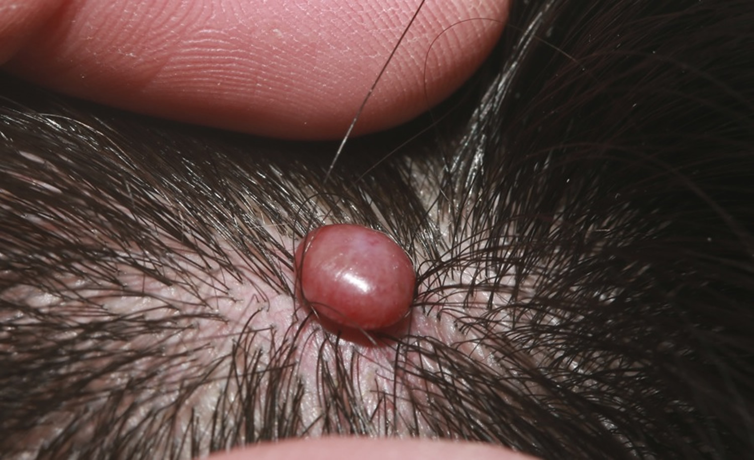
Cherry Angioma Scalp : Treating Anigoma Pure Dermatology - Cherry angiomas are red moles on your skin which contain an abnormal amount of blood vessels.
Get link
Facebook
X
Pinterest
Email
Other Apps
Cherry Angioma Scalp : Treating Anigoma Pure Dermatology - Cherry angiomas are red moles on your skin which contain an abnormal amount of blood vessels.. A hemangioma can occur anywhere on the body, but most commonly appears on the face, scalp, chest or back. They often appear on the torso of the body, though they can develop anywhere, including the arms, legs, chest, and even the scalp. Campbell de morgan spots, also known as cherry angiomas, are common, benign skin lesions of middle to older age, formed by proliferating, dilated capillaries and postcapillary venules. Cherry hemangioma in the scalp.pdf. The exact cause of cherry angioma is unknown but may be related to exposure to certain chemicals or changes in hormones.
Campbell de morgan spots, also known as cherry angiomas, are common, benign skin lesions of middle to older age, formed by proliferating, dilated capillaries and postcapillary venules. Hemangioma of skin and subcutaneous tissue. 75 % of people over age 70 have cherry angiomas on their body. Cherry angiomas or hemangiomas are little, bright red, pinpoint to match head sized, flat spots that over time, become raised, more dome shaped and ac. For small cherry angiomas, your dermatologist could use a device called a hyfrecator.
Cherry Angioma Removal Guide Younger Skin Guide from youngerskinguide.com It is the most common type of angioma. They often appear on the torso of the body, though they can develop anywhere, including the arms, legs, chest, and even the scalp. Cherry angiomas are small, red, harmless skin findings that occur commonly in older adults. It looks like a rubbery bump and is made up of extra blood vessels in the skin. Due to their bright appearance, your doctor or dermatologist should be able to tell you have a cherry angioma without needing to do any extensive testing. The underlying cause for the development of cherry angiomas is not understood, so nothing can be predicted about their prevention but rest assured; For example, keller reported that of hundreds of cases of cherry angiomas, only 2 cases were located on the scalp. Treatment options for cherry angiomas include laser treatment, electrodesiccation for typical lesions, and surgical excision for larger lesions.
Cherry angiomas or hemangiomas are little, bright red, pinpoint to match head sized, flat spots that over time, become raised, more dome shaped and ac.
They are less common on the scalp. For example, keller reported that of hundreds of cases of cherry angiomas, only 2 cases were located on the scalp. Cherry hemangioma in the scalp * josé marcos pereira. The face, scalp, neck, arms and legs are also probable sites for the development of cherry angiomas. Cherry angioma (campbell de morgan's spot) cherry angiomas are acquired vascular lesions that occur in up to 50 percent of adults. Cherry hemangioma (ch) is an extremely frequent dermatosis with vascular origin involving more than 75% of the population over 70 years of age. On the other hand, pereira reported that a high frequency of cherry angiomas occurred on the scalp. They often appear on the torso of the body, though they can develop anywhere, including the arms, legs, chest, and even the scalp. Cherry angiomas are usually multiple. Cherry angiomas are benign growths made up of blood vessels, which give them their characteristic red color. They are common on the trunk and limbs especially with advancing age. It looks like a rubbery bump and is made up of extra blood vessels in the skin. For small cherry angiomas, your dermatologist could use a device called a hyfrecator.
Cherry angiomas are benign growths made up of blood vessels, which give them their characteristic red color. They are the most common kind of angioma, and increase with age, occurring in nearly all adults over 30 years. In pereira's report, about 72% of patients had cherry angiomas on the. They're also known as senile angiomas or campbell de morgan spots. Cherry angiomas are usually multiple.
Growing Scalp Nodule Mdedge Family Medicine from cdn.mdedge.com When thrombosed, it can appear black in colour until examined with a dermatoscope when the red or purple colour is more easily seen. Cherry hemangioma in the scalp * josé marcos pereira. All skin hemangiomas will be visible by six months of age.they may occur anywhere on the skin surface, but they are most common on the scalp, face and neck. Rarely, a cherry hemangioma lesion demonstrates a dark brown to an almost black color. A hemangioma can occur anywhere on the body, but most commonly appears on the face, scalp, chest or back. Cherry hemangioma in the scalp.pdf. If they are to be removed for cosmetic reasons, electrosurgery and cryosurgery can be used. Content may be subject to copyright.
Cherry hemangioma in the scalp * josé marcos pereira.
Cherry hemangioma (ch) is an extremely frequent dermatosis with vascular origin involving more than 75% of the population over 70 years of age. Cherry angiomas, also known as campbell de morgan spots or senile angiomas, are cherry red papules on the skin. Cherry angiomas are red moles on your skin which contain an abnormal amount of blood vessels. However, i would highly recommend you try these homemade remedies for the sake of your health. Cherry angiomas are small, red, harmless skin findings that occur commonly in older adults. Limbs, face scalp, neck, but rarely the hands. A hemangioma can occur anywhere on the body, but most commonly appears on the face, scalp, chest or back. Rarely, a cherry hemangioma lesion demonstrates a dark brown to an almost black color. Cherry hemangiomas may be found on any body location. They are clumps of overgrown cells derived from the inside of blood vessels, or vascular endothelium. They are the most common kind of angioma, and increase with age, occurring in nearly all adults over 30 years. These skin lesions are extremely common. Herein, we report a case of multiple cherry angiomas on the scalp, an uncommon location for cherry angiomas.
They are common on the trunk and limbs especially with advancing age. Cherry angiomas are asymptomatic and have no reported clinical consequences. Cherry hemangiomas may be found on any body location. The papules seldom appear on the hands and feet. The underlying cause for the development of cherry angiomas is not understood, so nothing can be predicted about their prevention but rest assured;
Hemangioma Hemangioma Of Skin Spine Liver Causes Treatment from healthjade.com 75 % of people over age 70 have cherry angiomas on their body. Cherry angiomas are red moles on your skin which contain an abnormal amount of blood vessels. Your derm will apply a numbing cream first, but the treatment might still hurt a bit, she says. When thrombosed, it can appear black in colour until examined with a dermatoscope when the red or purple colour is more easily seen. The exact cause of cherry angioma is unknown but may be related to exposure to certain chemicals or changes in hormones. They're also known as senile angiomas or campbell de morgan spots. Cherry hemangioma in the scalp * josé marcos pereira. Cherry angiomas are made up of groups of dilated capillaries that are evident on the skin.
Cherry angiomas are asymptomatic and have no reported clinical consequences.
If they are to be removed for cosmetic reasons, electrosurgery and cryosurgery can be used. For small cherry angiomas, your dermatologist could use a device called a hyfrecator. They're also known as senile angiomas or campbell de morgan spots. Your derm will apply a numbing cream first, but the treatment might still hurt a bit, she says. In pereira's report, about 72% of patients had cherry angiomas on the. Cherry hemangiomas may be found on any body location. More recently pulsed dye laser or intense pulsed light (ipl) treatment are also used. While most people form cherry angiomas on the abdomen and trunk, it's also possible for them develop on the shoulders, upper chest, scalp, face, neck and arms, especially with older age. Cherry hemangioma (ch) is an extremely frequent dermatosis with vascular origin involving more than 75% of the population over 70 years of age. They often appear on the torso of the body, though they can develop anywhere, including the arms, legs, chest, and even the scalp. They are common on the trunk and limbs especially with advancing age. Content may be subject to copyright. Cherry hemangioma in the scalp.pdf.
Cherry angiomas are benign growths of blood vessels cherry angioma. These skin lesions are extremely common.


Comments
Post a Comment